When considering the health and well-being of livestock and other large animals, parasites are a major concern for veterinarians and farmers alike. These tiny, often invisible invaders can cause a range of diseases and health problems, from mild discomfort to severe illness or even mortality. To effectively manage these threats, it’s crucial to understand how these parasites look under the microscope. This article will explore some of the most common large animal parasites found in Europe, offering a microscopic view of their distinctive features.
1. Haemonchus contortus (Barber’s Pole Worm)
Host: Sheep, goats, and cattle
Transmission: Ingestion of larvae from contaminated pasture
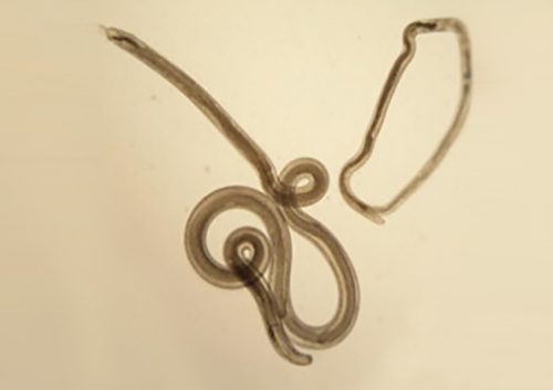
Haemonchus contortus, commonly known as the Barber’s Pole Worm, is one of the most notorious gastrointestinal parasites in livestock. It predominantly affects small ruminants such as sheep and goats but can also infect cattle. Haemonchus contortus are blood-sucking parasites that lay eggs in the abomasum of their host. The worm is large (up to 3 cm long) and identifiable by their distinct red and white spiral appearance, as a result of its blood filled intestinal tract, which resembles a barber’s pole.
Microscopic features:
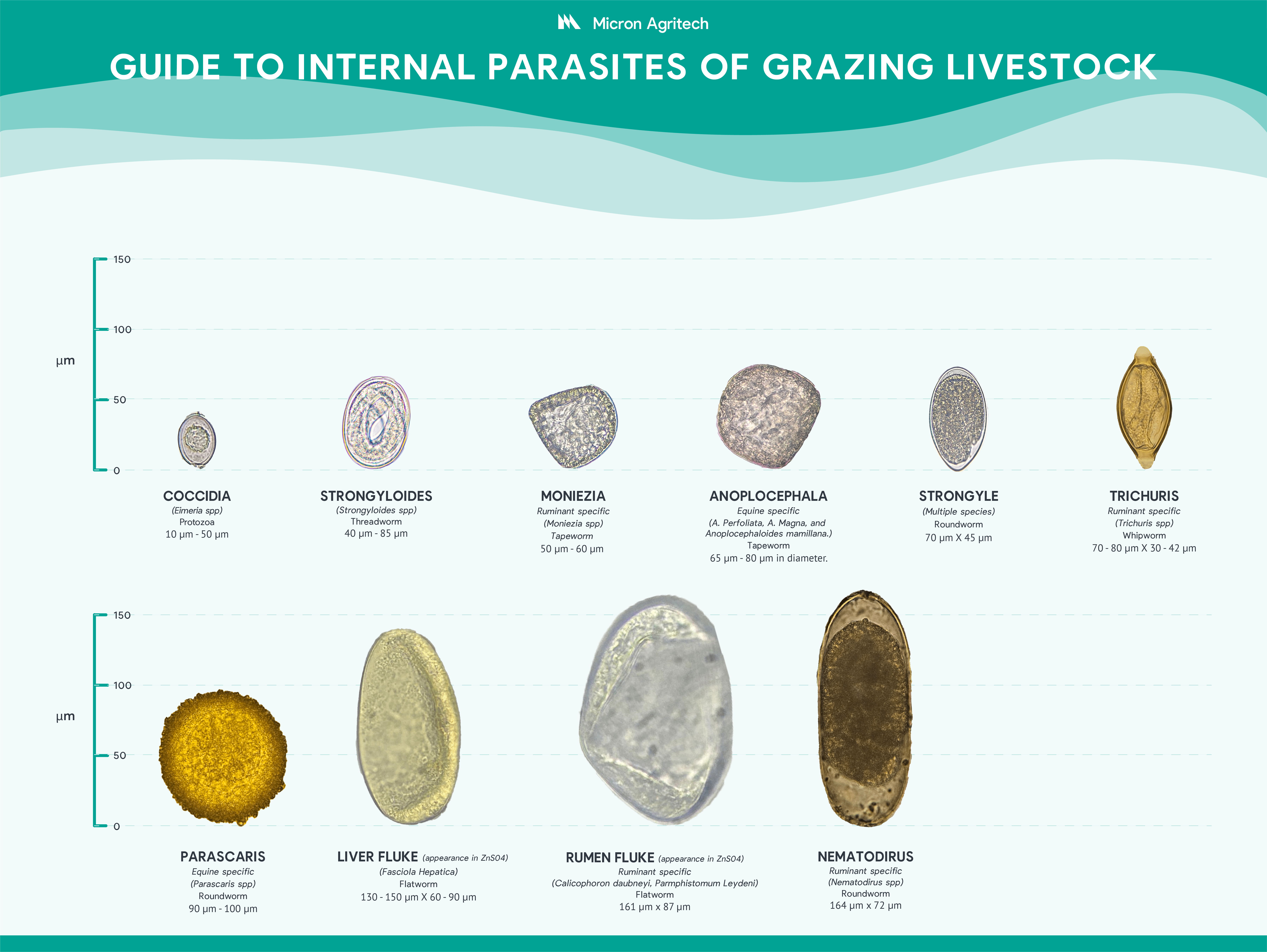
Strongyle
- The Haemonchus egg is oval and transparent, measuring about 70-85 microns in length.
- The larvae in the early stages (L1, L2) are also transparent, but as they develop into L3, they become more noticeable with their small, round bodies and characteristic movement.
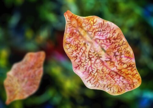
2. Fasciola hepatica (Liver Fluke)
Primary Host: Cattle, sheep, goats, and deer
Secondary Host: Mud snail
Transmission: Ingestion of cysts from contaminated water or vegetation
Liver flukes, particularly Fasciola hepatica, are widespread in Europe, especially in wetter regions. These parasitic flatworms can cause significant liver damage in their hosts, leading to symptoms such as weight loss, anemia, and jaundice. Under the microscope, Fasciola hepatica appears as a large, flat, leaf-shaped worm, typically measuring between 2 to 3 cm in length. The eggs of liver flukes are operculated (they have a lid) and oval, with lengths ranging from 130 to 150 microns.
Microscopic features:
- The eggs of Fasciola are easily distinguished due to their large size and operculated ends.
- Mature adult flukes can be seen under the microscope in faecal samples as flattened, reddish-brown structures, often with visible internal organs.
3. Trichostrongylus spp. (Hair Worms)
Host: Cattle, sheep, goats, and horses
Transmission: Ingestion of larvae from contaminated pasture
Trichostrongylus species are another common group of gastrointestinal worms that affect a wide variety of herbivores. These worms are usually found in the small intestine and cause symptoms such as lack of appetite, weight loss, malabsorption of proteins, and sometimes severe diarrhea. The adult worms are thin and threadlike, measuring about 1 cm in length. The eggs are relatively small and can be difficult to differentiate from other similar-sized nematodes.
Microscopic features:
Strongyle
- The eggs of Trichostrongylus species are smooth, oval, and typically measure around 70-90 microns.
- Larvae (L3 stage) are more easily identifiable due to their long, threadlike bodies and fast whip-like movement in wet mount preparations.
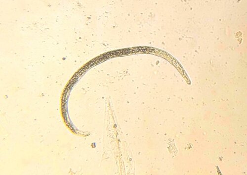
4. Strongylus vulgaris (Equine Strongyle)
Host: Horses
Transmission: Ingestion of larvae from contaminated pasture
Equine strongyles, particularly Strongylus vulgaris, are one of the most harmful internal parasites in horses. They are bloodsucking parasites and can cause significant damage to the intestines and lead to colic, anemia, and weight loss. The adult worms are red in colour and relatively large, measuring around 2 to 3 cm, the eggs and larvae are typically what is identifiable in faecal samples.
Microscopic features:
Strongyle
- Strongylus vulgaris eggs are slightly oval with a smooth thin shell, measuring around 80-100 microns in length
- The larvae at the L3 stage are more mobile and can be identified by their long, cylindrical bodies with a characteristic pointed tail.
5. Ostertagia ostertagi (Brown Stomach Worm)
Host: Cattle
Transmission: Ingestion of larvae from contaminated pasture
The Ostertagia ostertagi is one of the most common gastrointestinal parasites in cattle in Europe, particularly in temperate regions. It causes a disease known as “bovine ostertagiasis,” which can lead to severe digestive disturbances, such as loss in appetite, diarrhea, poor growth and in chronic cases, bottle jaw. The adult worm, found in the abomasum (the “true stomach” of cattle), is small, measuring about 1cm long, that borrows into the gastric glands.
Microscopic features:
Strongyle
- Eggs of Ostertagia are typically oval and thin-shelled, measuring about 80-100 microns.
- The larvae at the L3 stage are slightly larger and can be observed in faeces.

6. Moniezia spp. (Ruminant Tapeworm)
Primary Host: Sheep, goats and cattle
Secondary Host: Orbatid mite
Transmission: Ingestion of infectious mites from contaminated pasture
Moneizia spp. is the tapeworm found in small intestines of ruminants. This parasitic infection can display as asymptomatic. However, young ruminants or individuals with heavy infection can present clinical symptoms, such as diarrhea, poor growth or anemia. Adult worms are large and can measure different lengths, species dependant, ranging from 50cm to 5m.
Microscopic features:
- The Moneizia eggs are quadrangular in shape , measuring about 60 to 75 micrometers in diameter.
- The larvae in the early stages (L2) develop in the intermediate host, which once ingested penetrate the intestinal wall of the infected animal.
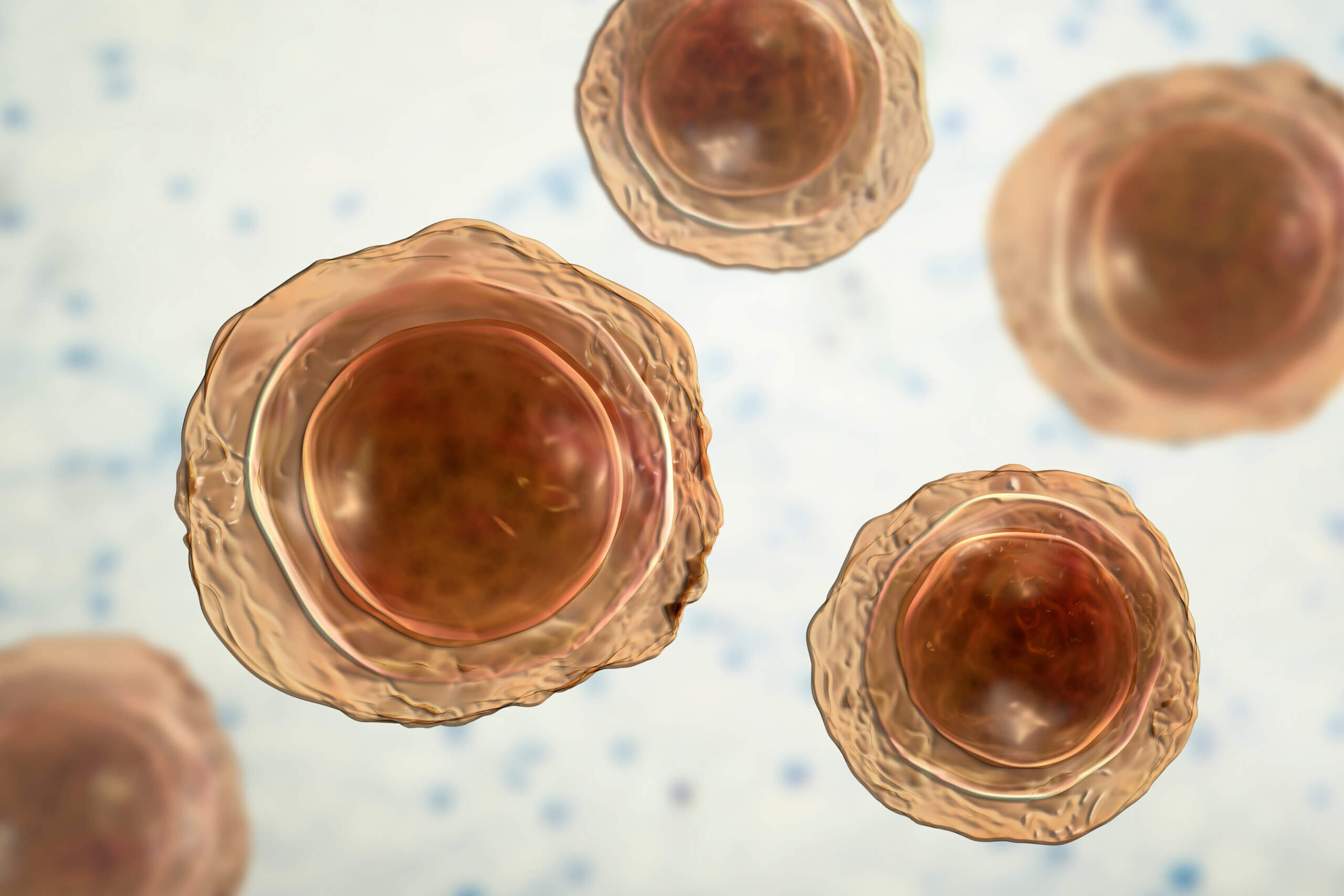
7. Eimeria spp. (Coccidia)
Host: Sheep, goats and cattle
Transmission: Ingestion of oocysts from contaminated pasture
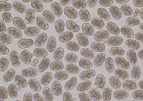
Eimeria spp. are protozoa commonly known as enteric coccidia that are found in the digestive tracts of infected animals. The Oocysts develop into sporulated oocysts in the environment and are ingested by grazing ruminants, resulting in penetration of the intestinal walls of infected animals. Clinical disease predominantly manifests in young animals. Coccidia are prolific and during their lifecycle of sporulation can produce millions of infectious sporulated oocytes. To avoid recurring disease due to the high oocyst counts graving management and reduced stress is required when managing coccidia.
Microscopic features:
- Oocysts have a thick resistant shell and can survive harsh conditions in the environment.
- Oocysts are ellipsoid in shame and range in length from 10 to 30 microns.
8.Trichuris spp. (Whipworm)
Host: Sheep, goats and cattle
Transmission: Ingestion of eggs from contaminated pasture and water
Trichuris spp.commonly referred to as Whipwom, is a genus of roundworm that infects the intestines of the host. Infections can lead to inflammation of the digestive tract, intestinal bleeding and reduced productivity in the animal. Adult worms can measure 30 to 50mm in length.
Microscopic features:
- Trichuris spp eggs are barrel shaped with a thick shell. These eggs have polar plug caps on each end measuring about 50 to 75 microns in length.
- The larvae stage hatch within the intestines of the animal and by burrowing into the caecum and colon.
9. Parascaris equorum
Host : Equine
Transmission: Ingestion of larvae from contaminate water or vegetation
Parascaris equorum is an ascarid roundworm found in equines. This parasitic infection can display as asymptomatic in light infections. However, young ruminants or individuals with heavy infection can present clinical symptoms, such as depressed growth development in foals, diarrhea and discomfort. The infective stage for horses is the egg containing a second-stage larva. Adult worms can measure up to 50 cm in length
Microscopic features:
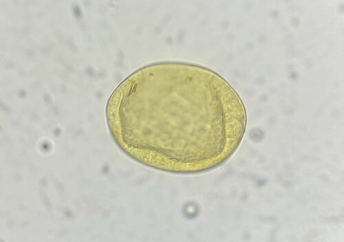
- Parascaris equorum eggs are brownish coloured measuring 90 to 100 µm in diameter.
- The eggs are nearly round in shape and have a thick rough outer shell.
10. Ascaris suum (Large Intestinal Worm)
Host: Pigs
Transmission: Ingestion of eggs from contaminated feed or water
Ascaris suum, or the large intestinal worm, is particularly common in pigs in Europe. Infected pigs often show signs of respiratory distress, poor growth, and digestive issues. The adult worms are amongst the largest nematodes, measuring up to 40 cm in length, and can be seen without a microscope. However, the eggs, which are oval and thick-shelled, are small and require a microscope to be properly examined.
Microscopic features:
- Ascaris eggs are distinctive due to their thick, rough outer shell and are about 50-75 microns in diameter.
- The eggs appear golden-brown and have a characteristic bumpy texture when viewed under a microscope
11. Nematodirus spp.
Host: Sheep, goats and cattle
Transmission: Ingestion of larvae from contaminated pasture
Nematoridus spp is a thread-necked roundworm that causes disease in the small intestine of ruminant animals. Nematodirus spp infections can result in poor nutrient transfer resulting in clinical signs of diarrhoea, dehydration and sudden weight loss. Adult worms can vary in length from 11 to 25 millimeters.
Microscopic features:
- Nematoridus spp, are large size eggs measuring 100 to 150 microns in length, inner material is distinctive with the large number of cells within the egg.
- The larvae in the later stages (L3) develop in the environment, which once ingested penetrate the intestinal wall of the infected animal.
12. Anoplocephala spp. (Equine Tapeworm)
Primary Host: Horses
Secondary Host: Orbatid Mites
Transmission: Ingestion of infected mites from contaminated pasture
Anoplocephala spp. is the tapeworm found in the small intestines of equine animals. This parasitic infection can display as asymptomatic. However, young ruminants or individuals with heavy infection can present clinical symptoms, such as diarrhea, anemia or colic . Adult worms are large and can measure different lengths, species dependant, ranging from 8 to 25 centimeters.
Microscopic features:
- Anoplocephala spp. Eggs can present as round or D-shaped with thick outer shell membranes measuring 65 to 80 microns in diameter.
- Anoplocephala spp. Larvae are passed through an intermediate host into the equine where it burrows into the small intestines of the animal.
13. Strongyloides spp. (Ruminant Threadworm)
Host: Cattle, sheep and goats
Transmission: Ingestion of is from contaminated pasture or direct skin penetration
Strongyloides spp.is a thread-like worm that is found in the intestinal tract of ruminants. Infection due to Strongyloides spp. result in symptoms such as diarrhea, weight loss, dermatitis and for young and immunocompromised animals these infections may be fatal. Adult female worms can be up to 3mm in length.
Microscopic features:
- Strongyloides spp. eggs are small oval shaped measuring from 50 to 80 µm in length.
- The eggs are identifiable due to their inner structure where the developing larvae can be visible.
Conclusion
Microscopy is a vital tool in the diagnosis and control of large animal parasites in Europe. By carefully examining faecal samples, veterinarians can identify the eggs, larvae, and sometimes even adult parasites that are causing issues in livestock and other large animals. Early detection and effective treatment of parasitic infections are key to maintaining healthy, productive animals. Understanding the microscopic features of these parasites allows for more precise identification, better treatment strategies, and ultimately a reduction in the impact of parasitic diseases on farming operations.
For farmers and veterinarians alike, keeping an eye on these microscopic invaders is essential for managing the health of large animals and safeguarding agricultural productivity.
References:
- Gordon, H. M., & Whitlock, H. V. (1939). A new technique for counting nematode eggs in sheep faeces. Journal of Council for Scientific and Industrial Research, 12(1), 50–52.
- Bowman, D. D. (2014). Georgis’ Parasitology for Veterinarians. Elsevier Health Sciences. This textbook covers identification and management of gastrointestinal parasites in large animals.
- Hansen, J., & Perry, B. (1994). The Epidemiology, Diagnosis, and Control of Helminth Parasites of Ruminants. International Livestock Centre for Africa.
- Rehbein, S., & Vervent, E. (2017). Parasites of Large Ruminants: The Role of the Veterinarian in Identifying and Managing Parasitic Diseases in European Livestock. Veterinary Parasitology, 245, 55-69.
- Levine, N. D. (1980). Nematode Parasites of Domestic Animals. Burleigh Dodds Science Publishing Ltd. This reference discusses the morphologic and lifecycle aspects of various nematodes including Haemonchus and Ostertagia.
- Relf, V. E., & Norris, J. M. (2011). The Importance of Regular Faecal Egg Counts in Livestock Health Management. British Veterinary Journal, 167(6), 250-255.
- MSD Veterinary Manual. Common gastrointestinal parasites of cattle – digestive system MSD Veterinary Manual. Available at: https://www.msdvetmanual.com/digestive-system/gastrointestinal-parasites-of-ruminants/common-gastrointestinal-parasites-of-cattle.
- Chemvet Australia (2024). Barber’s pole worm image (Figure 2). https://www.chemvet.com.au/barbers-pole-worm-haemonchus-contortus/https://www.chemvet.com.au/app/uploads/2024/10/B-Pole-Article-Thumbnail-300×300.png
- Bowman, D. D. (2014). Georgis’ Parasitology for Veterinarians (10th ed.). Elsevier.
- This textbook offers comprehensive coverage of parasitic infections in animals, including strongyloidosis and trichuriasis in ruminants. It discusses both the biology and impact of these parasites in livestock.
- Urquhart, G. M., Armour, J., Duncan, J. L., Dunn, A. M., & Jennings, F. W. (2013). Veterinary Parasitology (2nd ed.). Blackwell Publishing.
- A foundational text for veterinary students and professionals, this book covers the parasitic species that affect livestock, including the identification and control of Strongyloides and Trichuris.
- Scott, M. E., & McDougald, L. R. (2012). Veterinary Helminthology (2nd ed.). Blackwell Publishing.
- This reference provides in-depth information on nematode infections in livestock, with a special focus on gastrointestinal nematodes such as Strongyloides and Trichuris.
- Klein, M. L., & David, L. E. (2020). Diagnosis and Control of Helminth Parasites of Ruminants. Veterinary Clinics of North America: Food Animal Practice, 36(1), 99-112.
- This article provides a review of diagnostic techniques and control measures for helminth parasites in ruminants, including Strongyloides and Trichuris.
- Vercruysse, J., & Charlier, J. (2014). Control of Parasitic Helminths in Livestock. Parasitology, 141(S1), 124-133.
- A research article that covers the control and management of helminths, including Strongyloides and Trichuris, in large animals.
- Slocombe, J. O. D., & Power, K. F. (2016). Parasitic Diseases of Ruminants: A Field Guide for Practitioners. Elsevier.
- Provides practical guidance on the identification and treatment of common parasitic diseases in ruminants, with detailed sections on gastrointestinal nematodes like Trichuris and Strongyloides.
- Pritchard, G. C., & Holland, M. T. (2022). Gastrointestinal Nematodes in Ruminants: Diagnosis, Treatment, and Control. Veterinary Parasitology, 295, 12-23.
- An up-to-date review on the diagnostics and treatment protocols for gastrointestinal nematodes in ruminants, with particular focus on Strongyloides and Trichuris.
- Sykes, A. R. (2016). Helminth Parasites of Sheep and Cattle: A Review of Current Knowledge and Control Options. In: Helminth Infections in Ruminants: Control and Treatment. Springer.
- This chapter reviews key parasitic species and modern control strategies, with insights into Strongyloides and Trichuris infections in ruminants.
- Fossum, T. W. (2007). Small Animal Surgery (3rd ed.). Mosby.
- Although focused on small animals, this book provides useful insights into diagnosing and treating parasitic infections in large animals as well, including those caused by nematodes like Trichuris and Strongyloides.
- World Organisation for Animal Health (OIE). (2020). Manual of Diagnostic Tests and Vaccines for Terrestrial Animals.
- A resource for veterinary professionals, which includes diagnostic methods for a wide range of parasites, including Strongyloides and Trichuris.
- European Food Safety Authority (EFSA): Provides data and reports on animal health, including parasitic infestations in livestock.
The Merck Veterinary Manual (13th edition): Available online, this is a great resource for both common and rare parasitic infections in large animals.